Sleep and Aging: Why Pay More Attention to Sleep as We Get Older? Nature Aging Reveals New Changes in Brain Circulation
When it comes to sleep and aging, getting older may change more than just how long we sleep. This article explains a human study published in Nature Aging: older adults had weaker cerebrospinal fluid flow waves during non-rapid eye movement sleep, associated with reduced neural slow waves and changes in cerebrovascular responses. We will explore how the researchers observed these changes and what they mean for understanding brain aging.

We often find that sleep seems to become more difficult as we get older. When we are young, we may fall asleep as soon as our head hits the pillow. By middle and older age, however, we are more likely to wake up during the night, and deep sleep gradually decreases. [2]
We usually judge how well we slept by how long we slept and how alert we feel the next day. But sleep's effects on the brain may be far more complex than we imagine. Even after we fall asleep, a range of physiological activities continues inside the brain, including rhythmic neuronal activity, changes in cerebral blood vessels, and cerebrospinal fluid flow.
A human study published in Nature Aging on November 13, 2026, found that cerebrospinal fluid flow waves during sleep become markedly weaker with age. This change is associated not only with reduced slow-wave activity during sleep but also with diminished cerebrovascular responsiveness. [1]
This finding offers another perspective on the relationship between sleep and brain aging: getting older may change not just how long we sleep, but also how the brain functions during sleep.
What Happens Inside the Brain During Sleep?
The brain does not stop working when we fall asleep. Instead, it enters a physiological state that differs from wakefulness. During non-rapid eye movement (NREM) sleep in particular, neural activity, blood circulation, and cerebrospinal fluid flow in the brain undergo interconnected rhythmic changes. [1]
To understand these changes, we first need to look at three important physiological processes.
Neural Slow Waves: The Brain's Rhythmic Activity During Sleep
When we enter NREM sleep, the activity of large numbers of neurons in the cerebral cortex becomes more synchronized. Neurons periodically alternate between relatively active and relatively quiet states. These changes can produce slow-wave activity at approximately 0.5–4 Hz on an electroencephalogram. [1]
Slow waves are usually more prominent during deep sleep (slow-wave sleep), but their significance goes beyond helping researchers identify sleep stages. Neural activity also affects cerebral blood vessels through mechanisms such as neurovascular coupling, producing corresponding changes in cerebral hemodynamics. [1] [3]
Neural slow waves during sleep are therefore more than a sign that the brain is resting. They may also be the starting point for a series of physiological responses that regulate fluid movement within the brain.
Cerebral Blood Vessels: How Does Neural Activity Drive Fluid Movement in the Brain?
The brain is surrounded by a rigid skull. The space inside the skull is relatively fixed and is occupied mainly by brain tissue, blood, and cerebrospinal fluid.
When cerebral blood vessels dilate or constrict, blood volume inside the skull changes as well. Because space within the skull is limited, these changes in blood volume drive corresponding displacements of cerebrospinal fluid. [1]
During NREM sleep, neural slow waves can contribute to relatively slow oscillations in cerebral hemodynamics through neurovascular regulatory mechanisms. These changes, in turn, are closely related to the back-and-forth movement of cerebrospinal fluid. [1]
In other words, neural activity, changes in cerebral blood vessels, and cerebrospinal fluid flow are not three entirely independent processes. They are physiological systems that can interact and change together.
Cerebrospinal Fluid: Why Does Its Flow Deserve Attention?
Cerebrospinal Fluid (CSF) is a clear fluid found in the ventricular system and surrounding the brain and spinal cord.
It not only cushions external impacts and protects neural tissue but also helps maintain a stable environment inside the brain and participates in processes such as the transport of substances.
In recent years, researchers have increasingly focused on the exchange between cerebrospinal fluid and the fluid between brain cells, and on the role these fluids may play in transporting and clearing metabolic waste products from the brain. This is also an important area of glymphatic system research. [1]
Cerebrospinal fluid dynamics are part of the brain's normal physiology, and neural and vascular activity during sleep influences this process.
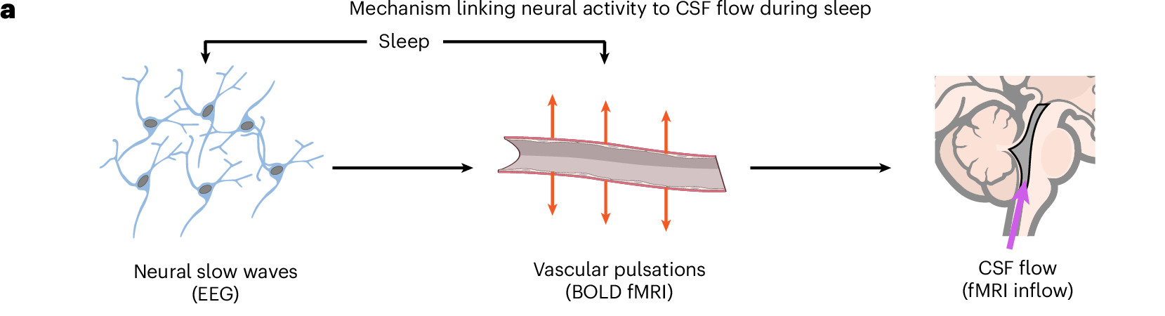
Figure 1: How Neural Slow Waves, Vascular Pulsations, and Cerebrospinal Fluid Flow Connect (Original Paper, Fig. 1a)
From left to right, the diagram shows neural slow waves, vascular pulsations, and cerebrospinal fluid flow. Neural activity during sleep affects cerebral blood vessels; changes in blood volume caused by vessel dilation and constriction then drive cerebrospinal fluid displacement. This figure illustrates how the three are connected. It does not measure the efficiency of waste clearance from the brain.
Adapted from: Bailes, S. M., et al., Nature Aging (2026), Fig. 1a. CC BY 4.0.
Why Does Aging Change Cerebrospinal Fluid Flow During Sleep?
To investigate whether aging affects this mechanism, the researchers analyzed nighttime sleep data from 72 healthy adults: 31 younger adults ages 18–39 and 41 older adults. [1]
The team used simultaneous electroencephalography (EEG) and fast functional magnetic resonance imaging (fMRI) to observe neural activity, changes in cerebral hemodynamics, and signals related to cerebrospinal fluid flow during participants' nighttime sleep. [1]
The results showed that low-frequency cerebrospinal fluid flow signals increased markedly when younger adults entered NREM sleep, while the increase was smaller in older adults.
Specifically, low-frequency cerebrospinal fluid signal power increased by an average of 3.82 dB from wakefulness to NREM sleep in the younger group, compared with just 1.15 dB in the older group. These values represent changes in signal power, not cerebrospinal fluid flow speed or percentages of waste clearance efficiency. [1]
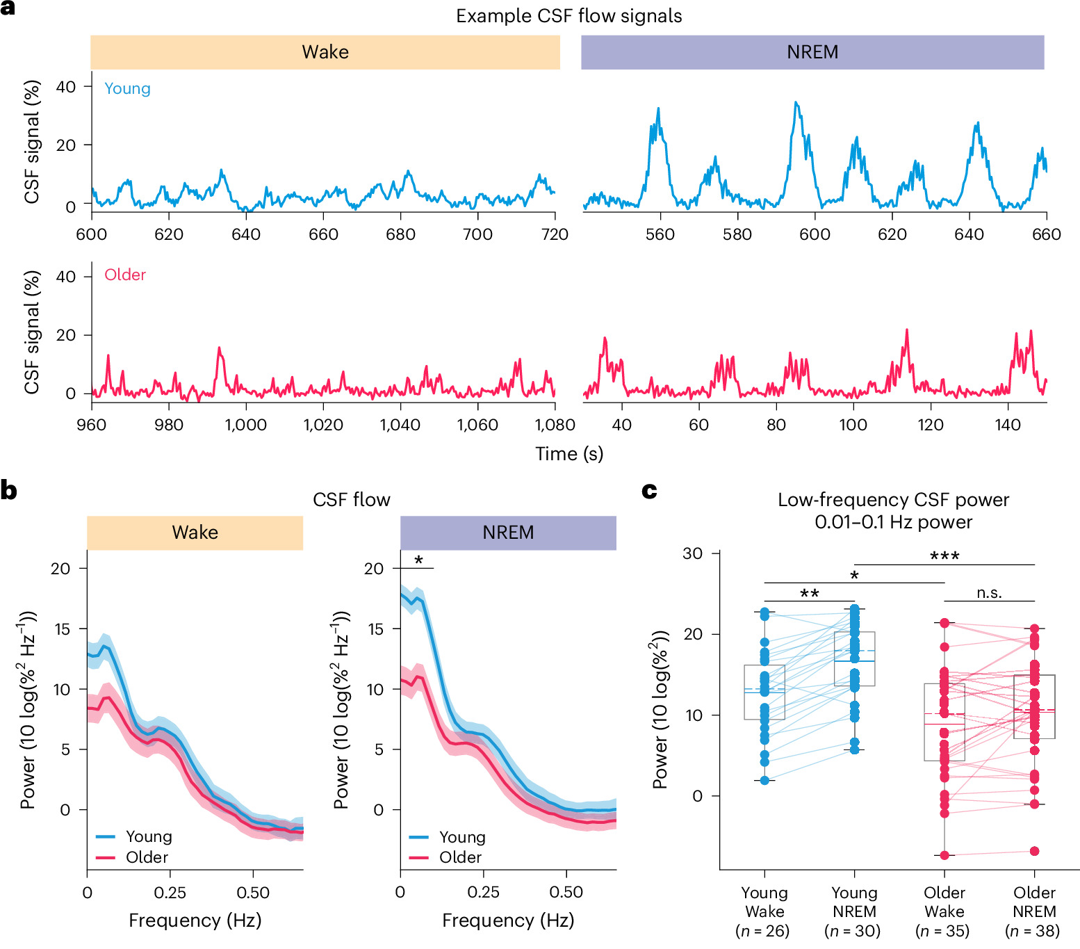
Figure 2: Differences in Cerebrospinal Fluid Signals During Sleep Between Younger and Older Adults (Original Paper, Fig. 2a–c)
Blue represents the younger group, and pink represents the older group. Panel a at the top shows signals from two representative participants: the cerebrospinal fluid signal fluctuates more noticeably when the younger participant enters NREM sleep. Panels b and c below show the group results: low-frequency signal power increased significantly from wakefulness to sleep in the younger group, while the increase in the older group did not reach statistical significance (n.s.). Signal power reflects the strength of fluctuations, not the total volume of cerebrospinal fluid or the rate of waste clearance.
Adapted from: Bailes, S. M., et al., Nature Aging (2026), Fig. 2a–c. CC BY 4.0.
Of particular interest, the study found that this change may involve two different physiological pathways.
The First Pathway: Reduced Slow-Wave Activity During Sleep
Sleep architecture typically changes with age. Deep sleep and slow-wave activity, in particular, tend to decrease. [2] [3]
To learn more about general age-related patterns in deep sleep, you can read about how much deep sleep you need at different ages. The reference ranges should not be treated as targets that everyone must meet.
The study found that EEG slow-wave power during NREM sleep was markedly lower in older adults than in younger adults. People with weaker slow-wave activity also tended to have weaker cerebrospinal fluid flow signals. [1]
This suggests that a reduction in sleep slow waves may weaken the neural drive behind cyclical changes in cerebral blood vessels, which may in turn affect cerebrospinal fluid flow. [1]
However, reduced slow-wave activity does not mean that cerebrospinal fluid flow will necessarily decrease by the same proportion. The cerebrovascular system also plays a role in the steps linking neural signals to cerebrospinal fluid flow.
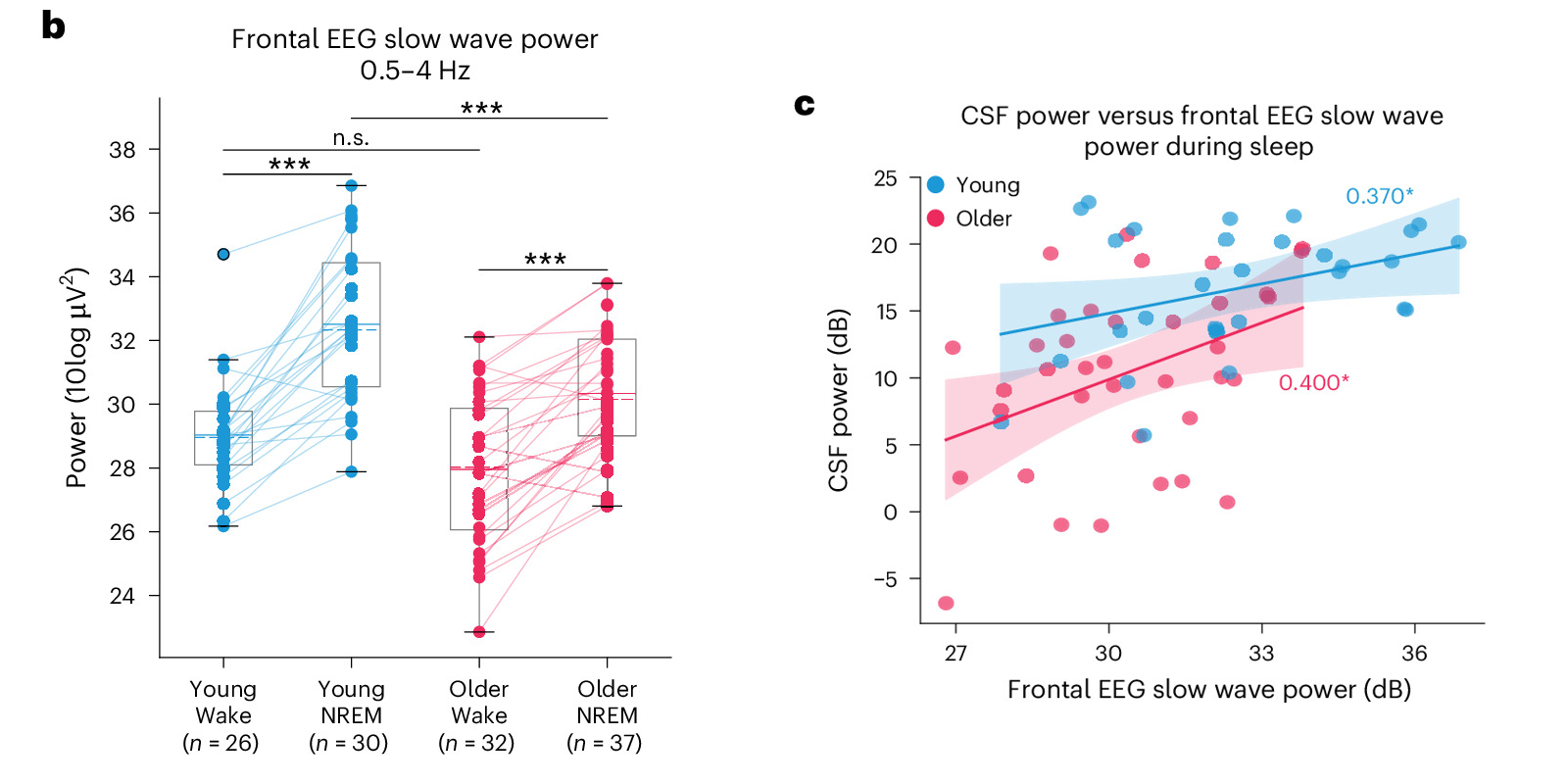
Figure 3: The Relationship Between Slow-Wave Activity and Cerebrospinal Fluid Signals (Original Paper, Fig. 4b, c)
Blue represents the younger group, and pink represents the older group. Panel b on the left shows that slow-wave power increased in both groups when they entered NREM sleep, but slow-wave power during sleep was lower in the older group. In panel c on the right, each point represents one participant: within each group, higher slow-wave power tended to accompany higher cerebrospinal fluid signal power. This shows an association, not that increasing slow waves will necessarily produce a proportional increase in cerebrospinal fluid flow.
Adapted from: Bailes, S. M., et al., Nature Aging (2026), Fig. 4b, c. CC BY 4.0.
The Second Pathway: Reduced Cerebrovascular Responsiveness
This is a particularly noteworthy finding from the study.
With age, cerebral blood vessels themselves may also change, including increased stiffness, reduced vascular reactivity, and changes in neurovascular coupling. [1]
This means that even if the brain can still generate neural slow waves, cerebral blood vessels may not respond as fully as they did at a younger age.
To investigate further, the researchers conducted visual stimulation and breath-holding experiments while participants were awake. These examined, respectively, hemodynamic responses to neural activity and the ability of cerebral blood vessels to respond to a physiological stimulus. [1]
The results showed that some cerebrovascular responses and the corresponding cerebrospinal fluid flow responses were weaker in older adults during these experiments. [1]
Age-related changes in cerebrospinal fluid flow may therefore reflect not only reduced sleep slow waves but also changes in cerebrovascular function itself.
Even if an older adult can still generate some sleep slow waves, cerebrospinal fluid flow during sleep may still be affected if cerebral blood vessels cannot respond effectively to those neural signals.
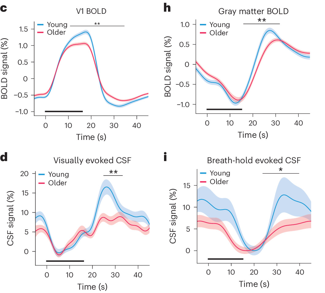
Figure 4: Vascular and Cerebrospinal Fluid Responses During Visual Stimulation and Breath-Holding Experiments (Original Paper, Fig. 6c, d, h, i)
Blue represents the younger group, and pink represents the older group. Panels c and d in the left column show the visual stimulation experiment, while panels h and i in the right column show the breath-holding experiment. The top row shows hemodynamic responses (BOLD), and the bottom row shows cerebrospinal fluid signals (CSF). In both experiments, responses were weaker in the older group during some time intervals; asterisks indicate significant differences between groups. These results support the explanation that changes in vascular responsiveness may contribute to weaker cerebrospinal fluid flow. Breath-holding was a research procedure, not a recommendation for improving sleep or cerebrospinal fluid flow.
Adapted from: Bailes, S. M., et al., Nature Aging (2026), Fig. 6c, d, h, i. CC BY 4.0.
Does This Mean Sleep Can Slow Brain Aging?
This question deserves discussion, but it requires a careful answer.
The study also found an association between weaker cerebrospinal fluid flow signals during sleep and lower frontal gray matter volume. [1] The frontal lobe is involved in important cognitive functions such as attention, working memory, decision-making, and behavioral control. This finding therefore offers new clues about the connection between sleep physiology and brain aging.
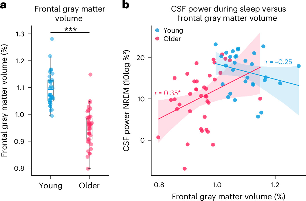
Figure 5: Cerebrospinal Fluid Signals and Frontal Gray Matter Volume: Association, Not Causation (Original Paper, Fig. 7)
Blue represents the younger group, and pink represents the older group. Panel a on the left shows that frontal gray matter volume, normalized to intracranial volume, was smaller in the older group. Panel b on the right shows that, within the older group, people with higher cerebrospinal fluid signal power during sleep tended to have greater relative frontal gray matter volume (r = 0.35); the correlation in the younger group was not significant. This is only an association. It does not prove that weaker cerebrospinal fluid flow causes brain atrophy or that increasing deep sleep can reverse brain aging.
Source: Bailes, S. M., et al., Nature Aging (2026), Fig. 7. CC BY 4.0.
That is because this study primarily compared physiological differences between age groups. It did not use a long-term sleep intervention to test whether brain aging could be slowed. [1]
The reality may be more complex. Changes in brain structure may affect sleep slow waves, declining cerebrovascular function may limit cerebrospinal fluid flow, and these factors may also be jointly influenced by other age-related physiological changes. [1] [2]
This study therefore does not directly prove that sleep counteracts aging. What it does reveal is that during normal aging, the physiological mechanisms that coordinate neural activity, cerebral hemodynamics, and cerebrospinal fluid flow during sleep may also gradually change.
How Should We Think About Sleep as We Get Older?
Discussions of sleep health have traditionally focused on how many hours we sleep each day, whether we wake up during the night, and whether we feel tired the next day. These measures still matter, but they do not fully reflect the physiological activity taking place inside the brain during sleep.
This study reminds us that sleep health involves more than sleep duration. It also includes neural activity, vascular function, and how they work together.
For middle-aged and older adults, maintaining a regular sleep schedule, allowing enough time for sleep, and paying attention to ongoing insomnia or problems such as sleep apnea remain important steps in managing health. [2] [4] Cardiovascular health also deserves attention, because cerebrovascular function may help regulate cerebrospinal fluid dynamics during sleep. [1]
To put these principles into practice, you can refer to seven practical adjustments to support good sleep and look for changes you can make to your schedule, caffeine timing, and bedtime habits.
Frequently Asked Questions (FAQ)
Is NREM Sleep in This Study the Same as Deep Sleep?
Not exactly. NREM sleep includes both lighter sleep stages and deep sleep, which usually refers to the N3 stage. The paper's findings about NREM sleep therefore cannot all be interpreted as changes that occur only during deep sleep. [3]
Does Less Deep Sleep With Age Mean the Brain Is Becoming Diseased?
You cannot draw that conclusion. Deep sleep and slow-wave activity change with age, and this study observed changes in cerebrospinal fluid signals in healthy older adults as well. These changes are not a basis for diagnosing brain disease. However, persistent insomnia, pronounced daytime sleepiness, or abnormal breathing during sleep should not simply be attributed to age. [1] [2]
Do Weaker Cerebrospinal Fluid Flow Waves Mean the Brain Is Less Effective at “Detoxing”?
This study cannot answer that directly. It measured signals related to cerebrospinal fluid flow and changes in cerebral hemodynamics, not the rate at which a particular metabolic waste product was cleared. Weaker flow waves therefore cannot be directly translated into a specific decrease in “detoxification efficiency.” [1]
Can Sleeping Longer Restore Cerebrospinal Fluid Flow in Older Adults?
That remains uncertain. This study did not test whether extending sleep or using other long-term interventions could reverse these changes. Getting enough sleep is still important, but these findings do not justify a promise that sleeping one extra hour will increase cerebrospinal fluid flow or reverse brain aging. [1]
If My Watch Shows Little Deep Sleep, Does That Mean My Cerebrospinal Fluid Flow Is Insufficient?
No. Consumer watches typically estimate sleep stages using signals such as movement and heart rate; they do not directly measure cerebrospinal fluid flow. This study used simultaneous EEG and fast functional magnetic resonance imaging. The number of deep sleep minutes on a watch cannot substitute for those measurements. [1] [5]
Final Thoughts: Focus on the Whole Night's Sleep, Not Just Deep Sleep Numbers
This study shows that the sleeping brain is far from quiet: neural activity, changes in cerebral blood vessels, and cerebrospinal fluid flow continue to work together, and that coordination changes with age. Weaker cerebrospinal fluid flow waves in older adults may involve both neural and vascular factors. They cannot be explained simply as “not enough deep sleep.” [1]
For sleep and brain health in everyday life, the more practical priorities are keeping a regular schedule, allowing enough time for sleep, and taking persistent insomnia, pronounced daytime sleepiness, or abnormal breathing during sleep seriously, rather than chasing a particular deep sleep score. [2] [4]
References
- [1] Bailes, S. M., et al. (2026). The aging human brain exhibits reduced hemodynamic and cerebrospinal fluid flow waves during sleep. Nature Aging. DOI: 10.1038/s43587-026-01270-4. DOI
- [2] Mander, B. A., Winer, J. R., & Walker, M. P. (2017). Sleep and Human Aging. Neuron, 94(1), 19–36. DOI: 10.1016/j.neuron.2017.02.004.
- [3] National Heart, Lung, and Blood Institute. (2022). How Sleep Works: Sleep Phases and Stages.
- [4] National Heart, Lung, and Blood Institute. (2022). Sleep Deprivation and Deficiency: Healthy Sleep Habits.
- [5] Chinoy, E. D., et al. (2021). Performance of seven consumer sleep-tracking devices compared with polysomnography. Sleep, 44(5), zsaa291. DOI: 10.1093/sleep/zsaa291.