Can't Feel Sleepy at Night? Here's How Sleep Drive Works
Why do you sometimes feel exhausted but not sleepy? Drawing on recent neuroscience research, this article explores how chemical signals and neural circuits in the brain regulate sleep pressure, explaining how sleepiness develops and offering a fresh scientific perspective on improving sleep.
Why do we feel a stronger urge to sleep the longer we stay awake? We generally think of sleepiness as something that gradually builds up during wakefulness. But how does the brain know how long we have been awake, and how does it turn a growing need for sleep into the act of sleeping?
Earlier research mainly emphasized the role of chemicals such as adenosine in the buildup of sleep pressure[4], while neuroscience research in recent years has found that specific neurons and neural circuits also help regulate sleep drive. These neurons not only respond to a growing need for sleep but can also actively influence whether sleep occurs. The interaction between chemical signals and neural circuits gives us a new understanding of how sleepiness develops.
The Brain Has Neurons Dedicated to Regulating Sleep Pressure
A mouse study published in Nature in 2026 found that specific neurons in two brain regions are closely linked to sleep drive: the anterior medial preoptic area (aMPO) and the median raphe (MR).[1]
The anterior medial preoptic area is located in the preoptic region of the hypothalamus and helps regulate sleep and wakefulness
The median raphe is located in the brainstem and contains several types of neurons with different functions.
The researchers found that some neurons in these two brain regions respond noticeably to sleep deprivation, with changes in their activity related to time spent awake and the need for sleep.
This indicates that the brain does not rely solely on changes in chemicals to regulate sleepiness. It also has specific groups of neurons that can respond to sleep pressure and change sleep behavior.
Brain Region Activity Associated With Wakefulness and Sleep Deprivation (Mice)
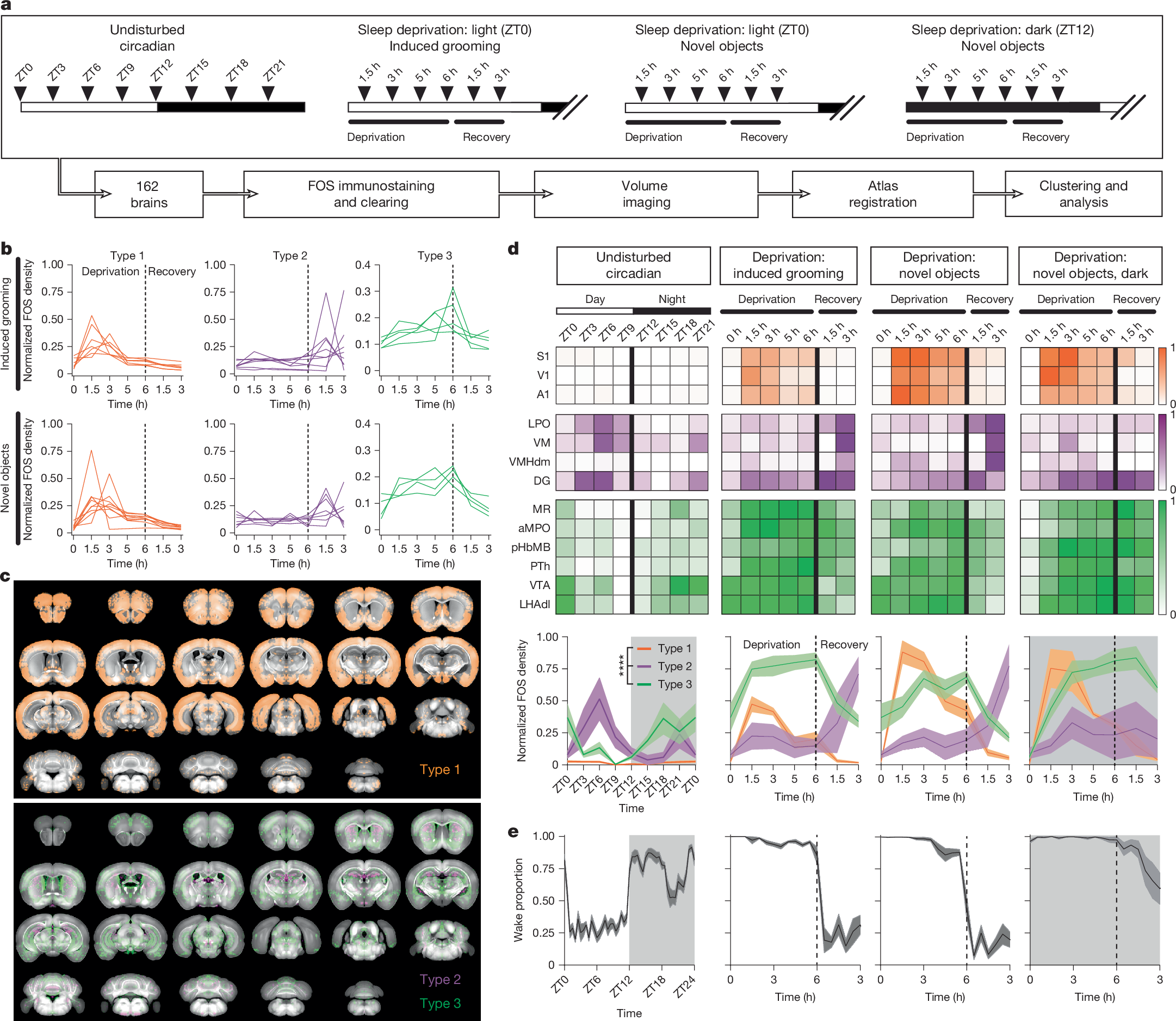
Focus on d and e: in d, MR and aMPO, shown in green, fall into the wakefulness-related class 3 response category. Viewed alongside the proportion of time spent awake in e, the corresponding FOS signals increase during sustained wakefulness/sleep deprivation and decrease during recovery sleep.
Source: Joo et al., Nature (2026), Fig. 1.
Why Do These Neurons Become More Active the Longer We Stay Awake?
The researchers observed a characteristic of these sleep-pressure-related neurons: as time spent awake increases, signals associated with their activity gradually strengthen; after sleep begins and recovery occurs, those signals gradually weaken.
This pattern appears not only in experiments involving imposed sleep deprivation but also during mice's normal day-night activity cycles. Mice are generally more active at night, when activity levels in the relevant neurons are higher. During the day, as sleep increases, those activity levels gradually decline. When researchers deliberately extend the time mice spend awake, these neurons also show more pronounced activation. This indicates that the changes are related not only to the time of day but also to the need for sleep that develops during sustained wakefulness.
More importantly, these neurons do not merely reflect sleep pressure passively. When researchers experimentally activated them, mice showed changes resembling recovery sleep, including longer sleep duration and greater sleep depth, even if they had not previously been sleep-deprived. Conversely, inhibiting these neurons reduced sleep and weakened the compensatory sleep response after sleep loss.[1]
Activating the Relevant Neurons Can Increase Sleep Amount and Slow-Wave Activity (Mice)
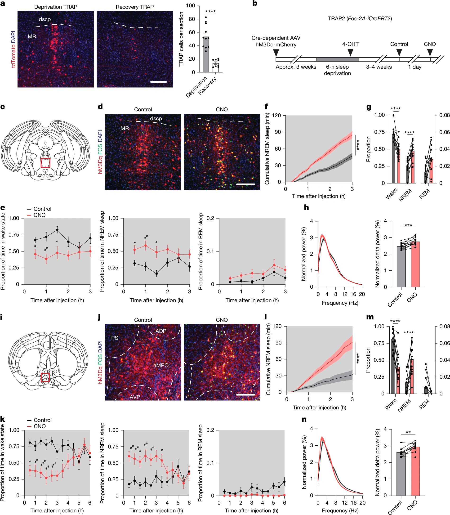
Focus on e–h (MR) and k–n (aMPO): red represents the CNO chemogenetic activation condition, and black represents the control. Activating cells tagged during sleep deprivation increased NREM sleep and δ-wave power, supporting the point in the text that active stimulation can induce changes resembling recovery sleep. δ-wave power is a measure of sleep intensity.
Source: Joo et al., Nature (2026), Fig. 2.
This shows that these neurons not only respond to the need for sleep but can also actively influence sleep drive. Their increasing activity during wakefulness is not intended to maintain wakefulness; instead, it may gradually push the brain toward a state of needing sleep.[2]
How Do Different Types of Neurons Work Together to Regulate Sleep?
Sleep is not controlled by a single type of neuron alone. In the median raphe, researchers identified three types of neurons with different functions: GABAergic, serotonergic, and glutamatergic neurons.
GABAergic neurons use γ-aminobutyric acid (GABA) as their primary neurotransmitter. GABA generally has an inhibitory effect, reducing the excitability of target neurons. The study found that some GABAergic neurons in the median raphe become more excitable after sleep deprivation. Activating these neurons can increase non-rapid eye movement sleep in mice, indicating that they promote sleep.
Serotonergic neurons help regulate neural activity through serotonin. Although serotonin can have different effects in different brain regions, this study found that the relevant serotonergic neurons in the median raphe can also promote sleep. When researchers activated GABAergic and serotonergic neurons simultaneously, the sleep-promoting effect lasted longer than when either type was activated alone, indicating that the two types work together.
Glutamatergic neurons have a different function. Glutamate is generally an excitatory neurotransmitter, and the study found that specific glutamatergic neurons in the median raphe primarily promote wakefulness. Activating them increases time spent awake and reduces non-rapid eye movement sleep.
Thus, these three types of neurons are not all responsible for promoting sleep; they play different roles in regulating sleep and wakefulness. GABAergic and serotonergic neurons work together to promote sleep, while some glutamatergic neurons promote wakefulness. Through its connections with other brain regions, the median raphe can also influence activity in sleep-promoting and arousal systems. This division of roles and cooperation allows the brain to dynamically regulate sleep and wakefulness according to its current need for sleep.[1]
Distinct Roles and Cooperation Among MR Neurons of Different Neurotransmitter Types (Mice)
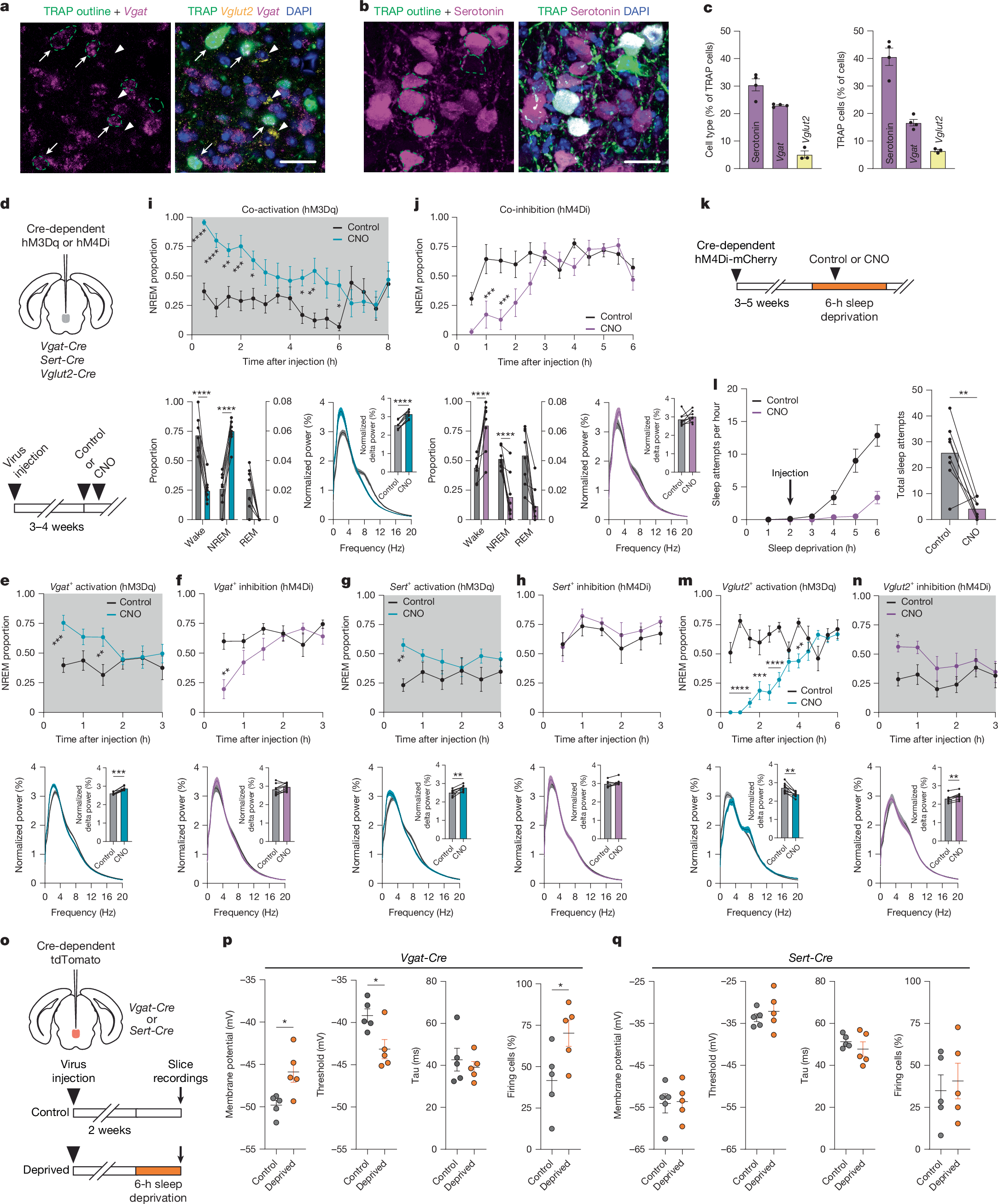
Focus on e, g, i, and m: activating either Vgat⁺ (GABAergic) or Sert⁺ (serotonergic) cells increases NREM sleep, while activating both types simultaneously produces a longer-lasting sleep-promoting effect. Activating Vglut2⁺ (glutamatergic) cells reduces NREM sleep. Panel p shows changes in the excitability of GABAergic cells after sleep deprivation.
Source: Joo et al., Nature (2026), Fig. 4.
A Closer Look: How Do Neurons “Record” Sleep Pressure?
The research discussed above tells us that specific neurons can respond to sleep pressure and work together to promote sleep. But another question remains: as time spent awake increases, why can the need for sleep continue to build and persist for a while?
One possible explanation is synaptic plasticity: the strength of connections between neurons can change with activity, and those changes can persist.
A fruit fly study published in Cell in 2016 found that prolonged wakefulness causes changes within specific neurons, mainly involving three aspects[3]:
Increased calcium ion concentrations: Calcium ions are important signals within cells. They can trigger a series of biochemical reactions that affect neuronal excitability and synaptic function.
Increased NMDA receptors: These receptors help transmit information between neurons and can help regulate synaptic strength through signals such as calcium ions.
Stronger synaptic connections: These intracellular changes are associated with synaptic plasticity, producing relatively lasting changes in the function of the relevant neural circuits.
Increased Sleep Need Is Accompanied by Elevated Calcium Signals in R2 Neurons (Fruit Flies)
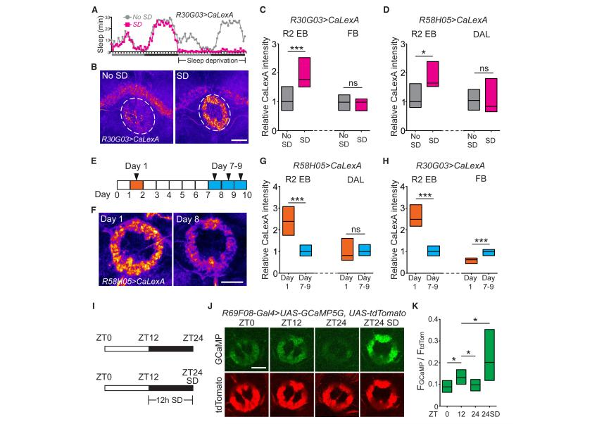
Focus on I–K: J shows the calcium indicator GCaMP and the reference signal tdTomato, while K shows the normalized quantitative results. The calcium-related fluorescence signal under the sleep-deprivation condition (ZT24 SD) is higher than under the non-deprived condition at the same time point (ZT24). SD stands for sleep deprivation. This result corresponds to the “increased calcium ion concentrations” described above.
Source: Liu et al., Cell 165, 1347–1360 (2016), Fig. 5.
NMDA Receptor-Dependent Plasticity and Sleep Drive (Fruit Flies)
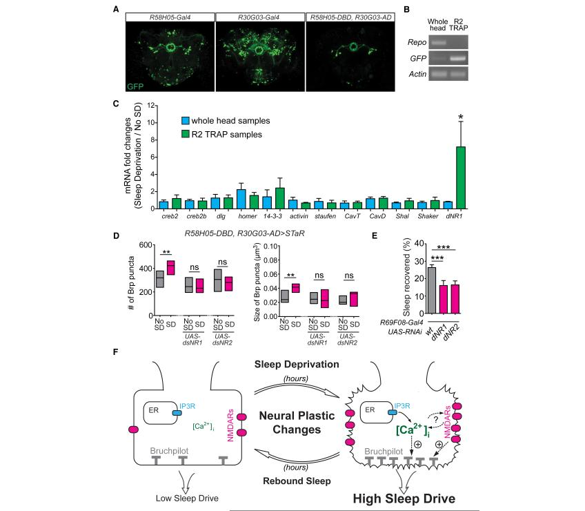
Panel C shows upregulation of dNR1 subunit mRNA in R2 cells after sleep deprivation (not a direct measurement of the number of receptor proteins). Panel D shows an increase in the number and size of puncta of the synaptic active zone marker BRP, a change that is blocked by knocking down NMDA receptor subunits. Panel E shows that the corresponding knockdown reduces compensatory sleep. BRP is a structural indicator of synaptic strength.
Source: Liu et al., Cell 165, 1347–1360 (2016), Fig. 7.
In simple terms, as time spent awake increases, calcium ion signals and NMDA receptors within the relevant neurons help regulate synaptic function, gradually strengthening the connections between neurons. This strengthening does not disappear as soon as a single episode of neural activity ends. Instead, it can last for a while, making the relevant neural circuits easier to activate. Prolonged wakefulness not only changes neuronal activity but may also leave lasting changes in the connections between neurons. Synaptic plasticity acts like a recording mechanism, allowing the brain to turn past wakefulness into a persistent drive to sleep. [3]
Final Thoughts
Sleepiness is not a switch that automatically flips on at bedtime. As time spent awake increases, signals related to sleep need change in the brain, specific neurons help promote sleep, and the plasticity of nerve cells may allow that need to persist. These animal studies give us a clearer view of the biological mechanisms behind sleep drive. For more on falling asleep, see how long it takes to fall asleep.
References
- [1] Joo, W., et al. (2026). Wake-activated neuronal populations that regulate sleep drive. Nature. DOI: 10.1038/s41586-026-10928-3. Figures reproduced under CC BY 4.0.
- [2] Luff, C. E., Ly, S. L., & de Lecea, L. (2026). Deciphering sleep drive in the brain: new circuit-level insights. Cell Research. DOI: 10.1038/s41422-026-01317-6. (Research Highlight: an interpretation of the research in reference [1], not an independent experiment.)
- [3] Liu, S., et al. (2016). Sleep Drive Is Encoded by Neural Plastic Changes in a Dedicated Circuit. Cell, 165, 1347–1360. DOI: 10.1016/j.cell.2016.04.013.
- [4] National Heart, Lung, and Blood Institute (NHLBI). (2022). How Sleep Works: Your Sleep/Wake Cycle. (Background information on sleep homeostasis and adenosine.)